








ChondroFiller® vs
Microfracture
Microfracture is a long-established arthroscopic technique for small cartilage defects. ChondroFiller® is a non-surgical scaffold injection that delivers cartilage regeneration without bone-marrow stimulation or arthroscopy. Here is the honest comparison.
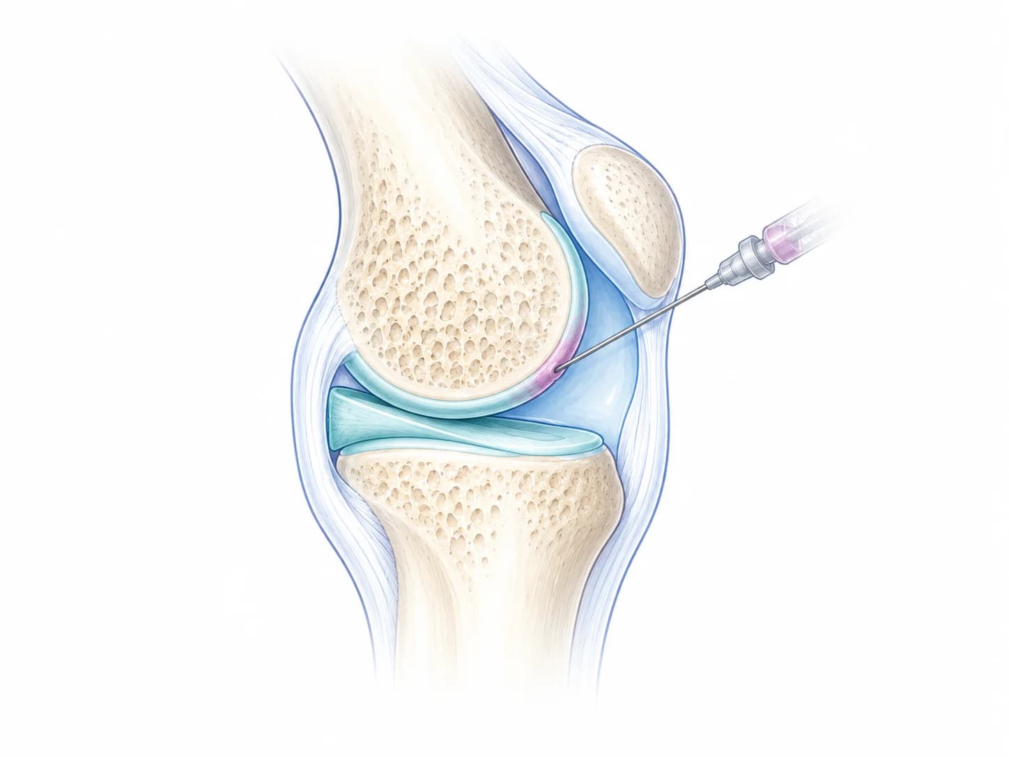
- Collagen scaffold
- Ultrasound-guided injection
- Coats the worn surface
ChondroFiller®
A collagen scaffold injected under ultrasound guidance. It sets in minutes and coats the worn cartilage so your own cells can lay down new tissue.
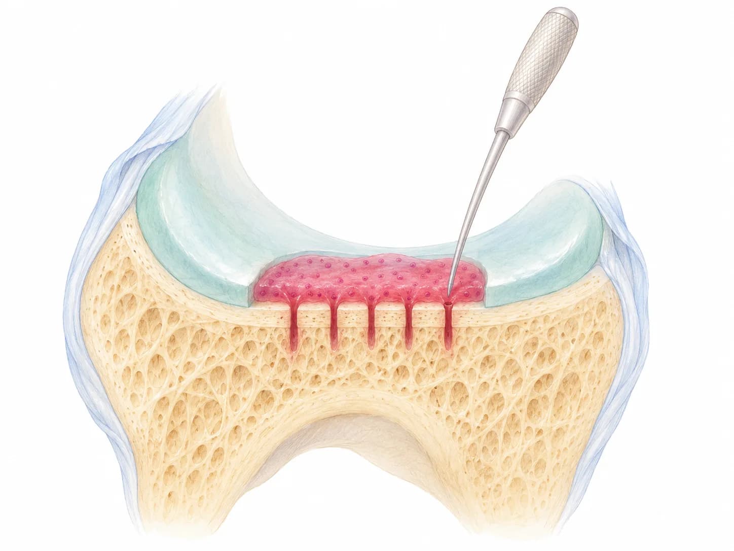
- Defect cleared
- Small holes in the bone
- Marrow forms repair tissue
Microfracture
Keyhole surgery that makes small holes in the bone beneath a defect so marrow can form repair tissue.
Quick answer
ChondroFiller® supports natural cartilage restoration without surgery; microfracture is a keyhole operation that stimulates the bone marrow to fill the defect with fibrocartilage. Microfracture has a long evidence base, particularly in younger active patients with small defects, but the repair tissue it produces is fibrocartilage, which can deteriorate after three to five years. ChondroFiller® aims to deliver more hyaline-like tissue and avoids drilling the subchondral bone. It has no upper age limit and no defect-size limit on the injection, and it coats the cartilage surface across the joint in a single delivery. The choice is made at consultation with MRI review.
How it works
ChondroFiller® vs Microfracture: the key difference
Microfracture works by deliberately damaging the subchondral bone: the surgeon makes small holes through the bone plate so marrow cells leak into the defect and form a clot, which matures into fibrocartilage. ChondroFiller® instead places a sterile collagen scaffold directly into the mapped defect under ultrasound guidance; the patient’s own cells then migrate into the scaffold and form new cartilage matrix over six to twelve months. Different mechanism, different recovery, different repair tissue.
How each treatment works
ChondroFiller®
ChondroFiller® is a sterile type I collagen scaffold. At London Cartilage Clinic it is injected as a liquid into the joint under ultrasound guidance, where it sets within minutes and coats the worn cartilage at the load-bearing surfaces, from a contained focal defect to diffuse surface wear.
The patient’s own cells then migrate into the scaffold and lay down new cartilage matrix over six to twelve months. The subchondral bone is left alone: ChondroFiller® does not stimulate the marrow or alter the bone plate.
Microfracture
Microfracture is an arthroscopic procedure. The surgeon prepares the cartilage defect and then makes a series of small holes (microfractures) through the subchondral bone plate using an awl or drill.
Bone marrow leaks into the defect and forms a blood clot containing stem cells; over the following months this matures into fibrocartilage, repair tissue rich in type I collagen rather than the type II collagen found in native hyaline cartilage.
Looking ahead
What can you expect over time?
ChondroFiller®
Tissue formation over 6–12 months
Microfracture
Good early relief, often declines after 3–5 years
Read the comparison in context
Your treatment plan
Number of injections
ChondroFiller®
Typically one injection course, with the box quantity (one, two or three) decided from MRI. ChondroFiller® can be repeated if a separate new defect develops; the original implanted tissue is intended to be durable.
Microfracture
Microfracture is a one-off arthroscopic operation. Revision or conversion to a different cartilage procedure (MACI, autologous chondrocyte implantation, or a scaffold-based treatment) is sometimes needed years later if the fibrocartilage deteriorates.
The treatment journeys
ChondroFiller® pathway
- 1
Imaging review with Professor Lee
At consultation your MRI is reviewed to confirm the defect is suitable and to set the number of boxes. - 2
Injection under ultrasound
The gel is injected as a liquid under ultrasound guidance and local anaesthetic in an outpatient appointment of 30 to 45 minutes, and it sets within minutes. - 3
Back home, then a review call
You leave the same hour without a brace or crutches unless your medical team says otherwise, and the free telephone review comes at six weeks.
Microfracture pathway
- 1
Arthroscopic preparation
The surgeon prepares the cartilage defect at arthroscopy. - 2
Microfractures through the bone plate
Small holes are made through the subchondral bone plate using an awl or drill, so bone marrow leaks into the defect. - 3
Fibrocartilage forms
The marrow clot matures over the following months into fibrocartilage, with a rehabilitation protocol that often includes a period of restricted weight-bearing.
Care and precautions
Understanding the risks
BEFORE · DURING · AFTER
Sterile technique.
Image guidance.
Clear aftercare.
These are very different procedures with very different risk profiles. ChondroFiller® at London Cartilage Clinic is an ultrasound-guided outpatient injection delivered under full sterile technique with routine IV antibiotic cover. The most serious risk is joint infection, which is rare.
Microfracture is arthroscopic surgery: it carries the standard arthroscopy risks (infection, deep-vein thrombosis, anaesthetic complications) plus surgery-specific issues: subchondral bone overgrowth (intralesional osteophyte formation), bone marrow oedema and a rehabilitation protocol that often includes a period of restricted weight-bearing. See our injection safety and infection-prevention protocol for the ChondroFiller® injection-side detail.
Practical planning and safety
Questions to raise before either option
Ask about temporary soreness or swelling, the risk of joint infection, and anything specific to your health and current medicines. Ask who you contact if something does not feel right afterwards.
Who may suit ChondroFiller →Aftercare depends on the route
After the ChondroFiller injection there is no brace and no crutches unless your medical team specifies otherwise. Keyhole surgery follows the rehabilitation protocol set out by the manufacturer, with a brace for 48 hours and weight rebuilt over about six to eight weeks.
Read the injection aftercare guide →Your joint, your goals
Where does each option fit?
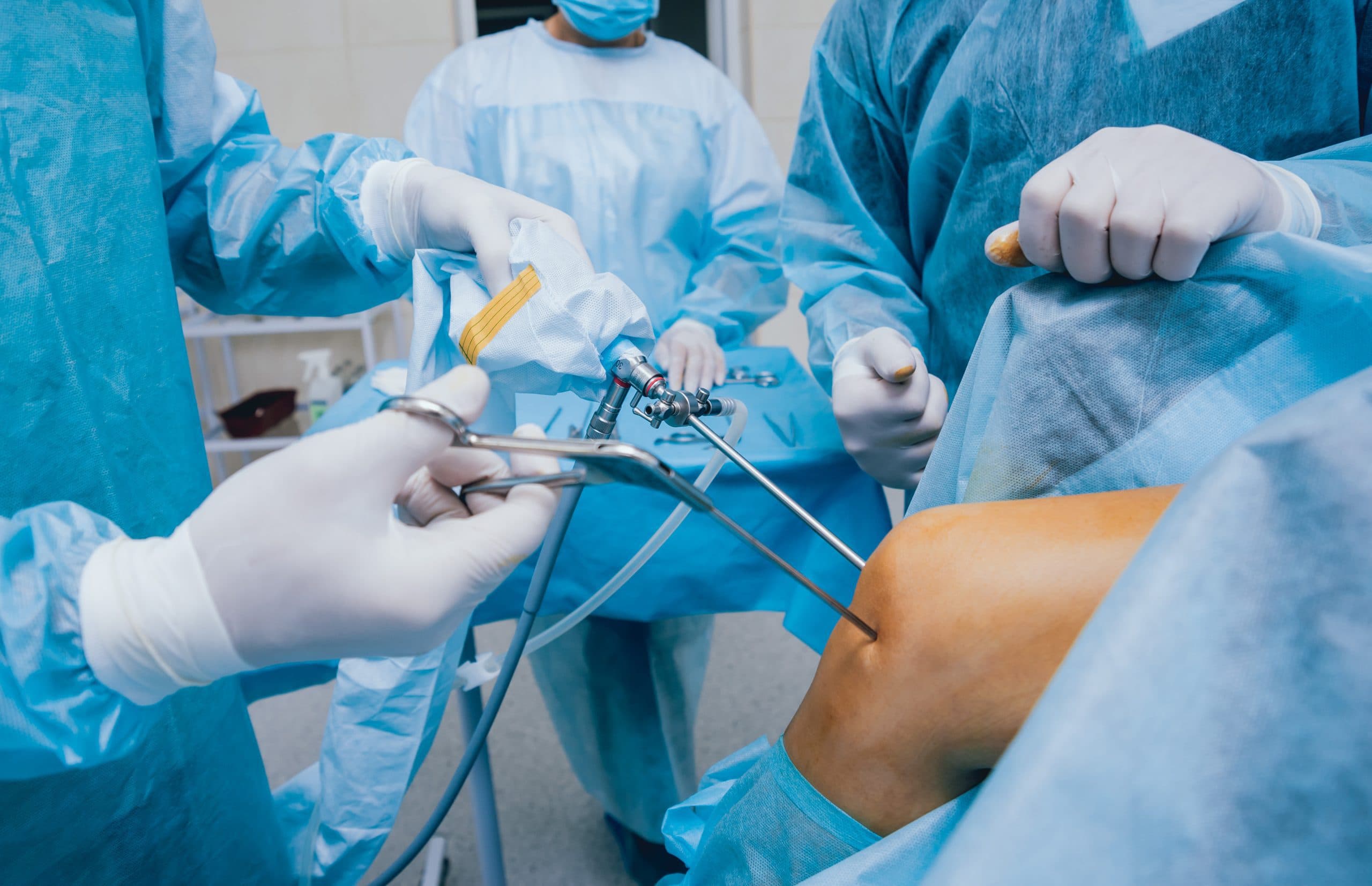
Microfracture is most defensible in younger, active patients with small contained cartilage defects (typically under 2 cm²), good bone quality and no significant generalised joint disease. In larger defects, older patients or joints with bone marrow oedema, modern cartilage protocols increasingly favour scaffold-based or cell-based options over microfracture.
ChondroFiller® is appropriate for cartilage wear in the joint, whether a contained focal defect or diffuse surface wear, with no upper age limit and no defect-size limit on the injection. It is less suitable where the subchondral bone is unstable or where the joint mechanics (instability, malalignment) are untreated. See who is suitable for ChondroFiller?
What would make the difference for you?
Questions worth taking to your consultation, so the discussion stays on your joint.
- 01What is causing my joint pain?The diagnosis and your imaging decide which treatments are worth discussing.
- 02Is there a defect, or wear across the joint?Ask how the pattern of cartilage damage shapes the recommendation.
- 03What improvement are we aiming for?Agree realistic goals for pain, function and timescale before you choose.
- 04What happens if I need more help later?Ask which options stay open if symptoms return or progress.
Planning the cost
Compare the complete care package
Ultrasound-guided injection
ChondroFiller®
From · one box
£3,000
Two boxes £5,500; three boxes £8,000. The consultation is charged separately.
Full ChondroFiller pricing →At London Cartilage Clinic, ChondroFiller® costs from £3,000 for one box, £5,500 for two and £8,000 for three. The price includes ultrasound-guided injection, IV antibiotic cover and six-week follow-up. The consultation is charged separately. See the ChondroFiller® cost guide.
Private microfracture in the UK is typically priced as an arthroscopic knee procedure, from around £6,000 to £9,000 depending on hospital, surgeon and anaesthetic. It is available on the NHS for clinically appropriate cases, though waiting times vary.
The headline numbers are similar; the more important comparison is the trade-off between a non-surgical scaffold and an arthroscopic bone-marrow-stimulation operation, and which one fits the defect on imaging.
Making a recommendation
When each option may make sense
ChondroFiller®
A discussion to have
- You have a focal cartilage defect on MRI and want a non-surgical pathway.
- You want to preserve the subchondral bone plate rather than have it deliberately perforated.
- You want a regenerative pathway that targets the defect with a scaffold rather than relying on marrow-stimulation to fill it with fibrocartilage.
Microfracture
A discussion to have
- A small, contained cartilage defect (typically under 2 cm²) in a young, active patient with otherwise healthy bone.
- A patient already booked for arthroscopy for another reason (loose body, meniscal repair) where the small defect can be addressed at the same time.
- Where surgery is the agreed plan and the surgeon judges marrow-stimulation to be the most appropriate cartilage step.
Choosing another path
When another approach may be better
ChondroFiller®
A reason to reconsider
- Joints where imaging review points to replacement; bone-on-bone wear on its own is not the deciding factor.
- Significant subchondral bone oedema or instability: the bone needs to be a stable base for new tissue.
- Very large or multi-zone defects where a surgical pathway (Liquid Cartilage™ or autologous chondrocyte implantation) is the better answer.
Microfracture
A reason to reconsider
- Larger defects (typically over 2–4 cm²), older patients or anyone with established bone marrow oedema: modern evidence increasingly favours scaffold- or cell-based options here.
- Patients who want to avoid arthroscopy when a reasonable non-surgical alternative (ChondroFiller® injection) exists.
- Patients who cannot tolerate a period of restricted weight-bearing as part of microfracture rehabilitation.
The bottom line
Microfracture is a legitimate cartilage-repair option with decades of clinical experience and a place in the modern surgical toolkit. ChondroFiller® offers a different approach: a scaffold-based, non-surgical pathway that preserves the subchondral bone and aims for more hyaline-like repair tissue. The right answer depends on defect size, location and the rest of the joint picture on MRI. We will tell you honestly when surgery is the better answer.
Common questions
01Is ChondroFiller® better than microfracture?
Better is the wrong word: they are different tools. Microfracture has decades of evidence and a clear role in small defects in young active patients. ChondroFiller® aims to produce more hyaline-like tissue without drilling the subchondral bone, and is delivered as an injection rather than a surgery. The right answer depends on the defect and the patient.
02Why does microfracture produce fibrocartilage and not hyaline cartilage?
The repair tissue that forms after microfracture comes from a marrow clot, and its natural maturation pathway produces fibrocartilage, a tissue rich in type I collagen, mechanically inferior to native hyaline cartilage (type II collagen). Hyaline cartilage is what scaffold- and cell-based techniques like ChondroFiller®, MACI and ACI are trying to recreate.
03I have already had microfracture and it has failed. Can I have ChondroFiller®?
Sometimes yes. Where the failed microfracture has left a contained defect with reasonable surrounding cartilage and a stable subchondral bone plate, ChondroFiller® is sometimes a reasonable next step. Where the bone plate is irregular or there is significant marrow oedema, a different procedure may be needed. We review the MRI before deciding.
04Does ChondroFiller® avoid the recovery time of microfracture?
ChondroFiller is one gel that can be placed two ways. By ultrasound-guided injection it is an outpatient appointment with no brace and no crutches unless your medical team specifies otherwise: about half your usual joint activity in weeks one and two, then a graded return by week six, with new tissue forming over six to twelve months. If the same gel is implanted by keyhole surgery, recovery is much slower, with a brace for 48 hours and partial weight-bearing for about six weeks. Microfracture typically requires a prescriptive protocol of its own because the subchondral bone has been perforated.
05Is microfracture available on the NHS?
Yes, microfracture is available on the NHS for clinically appropriate cartilage defects, though waiting times and surgeon preference vary. ChondroFiller® is not routinely funded by the NHS in the UK and is a private treatment at this time.
06Which has stronger evidence?
Microfracture has the longer publication history but the strength of that evidence has softened in recent years, particularly for defects larger than 2 cm². ChondroFiller® has published outcome data (IKDC, Harris Hip Score, MOCART MRI) and over 19,000 cases of clinical use. See the ChondroFiller® clinical evidence page.